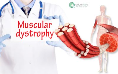
Regenerative Approach To Muscular Dystrophy Treatment
Muscular Dystrophy encompasses a group of genetic disorders characterized by progressive muscle weakness and degeneration. As the condition advances, it begins to affect muscles of vital organs, significantly impacting overall health and life expectancy. Although conventional therapies help slow disease progression and preserve muscle strength, recent breakthroughs in stem cell therapy have elevated treatment goals to a new level.
About Muscular Dystrophy (DP)
Muscular Dystrophy involves the gradual deterioration of muscle tissue due to mutations in genes - alterations in the DNA sequence. These mutations are typically inherited and affect genes responsible for producing proteins essential for muscle cell survival and function. The absence or malfunction of these proteins leads to muscle cell damage, triggering the body’s repair mechanisms. However, instead of regenerating healthy muscle cells, these processes often result in the accumulation of fat and connective tissue within the muscle, further impairing function.
Which muscles are primarily affected in muscular dystrophy?
In most cases, skeletal muscles are the first to be affected, leading to difficulty in movement of the limbs, especially the arms and legs. In certain types of muscular dystrophy, the weakness extends beyond skeletal muscles to include those of the heart and lungs, resulting in complications related to cardiac function and respiratory capacity.
How long do the patients with muscular dystrophies live?
The lifespan of individuals with muscular dystrophy varies widely depending on the specific type and its severity. For instance, Duchenne muscular dystrophy (DMD) is among the most severe forms, often limiting life expectancy to the early 20s. In contrast, Becker muscular dystrophy (BMD) tends to progress more slowly, allowing some individuals to live into their 50s or 60s. With ongoing advancements in medical science, improved healthcare access, and supportive therapies, many people with muscular dystrophy can maintain independence and a good quality of life for extended periods.
Does muscular dystrophy affect only in males?
Muscular dystrophy can affect both males and females. However, certain types—particularly those linked to mutations on the X chromosome are more prevalent in males due to the way these genetic traits are inherited.
While numerous treatments exist to manage muscular dystrophy, a definitive cure remains elusive. Researchers continue to explore various therapeutic avenues, with stem cell therapy emerging as one of the most promising. Its regenerative potential offers hope for restoring muscle function.
With the advent of technology and cutting-edge research, the treatment approach is gradually inclining towards customized solutions. Clinicians are increasingly tailoring treatment plans based on the specific type of muscular dystrophy, its severity, progression rate, and individual patient characteristics. This evolution in care strategy is paving the way for personalized stem cell treatment, offering more targeted and effective solutions for managing the disease.
Diagnosis
Early diagnosis of muscular dystrophy plays a pivotal role in enabling timely therapeutic intervention. Prompt treatment initiation can significantly delay the progression of muscle weakness and wasting. Therefore, beginning therapy at the earliest possible stage is strongly recommended to improve clinical outcomes and enhance overall quality of life.
Physical Examination: Clinicians often examine physical symptoms indicating muscular dystrophy such as enlarged calf muscle, weakness in lower limbs, support of arms while rising from a seated position, toe-walking, hip girdle muscle instability, etc.
Blood Tests: Muscle damage releases enzymes into blood such as creatinine kinase, aldolase, LDH, myoglobin, etc. Elevated levels of these biomarkers serve as indicators of muscular dystrophy. Additionally, some individuals may exhibit increased levels of liver enzymes like alanine transaminase (ALT) and aspartate aminotransferase (AST), which can also reflect muscle pathology.
Genetic Testing: Mutation analysis offers a definitive diagnosis. It employs techniques like PCR and sequencing to identify the mutations behind muscular dystrophy. With advancements in technology, genetic testing can now be performed using either blood samples or saliva, making the process more accessible and less invasive.
Imaging: Diagnostic imaging tools like MRI and CT scans are used to detect structural changes in muscle tissue associated with muscular dystrophy, such as increased intramuscular fat, reduce bone density, etc.
Muscle Biopsy: This technique involves collecting a small sample of muscle tissue for histological analysis. The biopsy reveals key features such as variation in muscle fiber size, presence of connective tissue, and fat infiltration, all of which are indicative of muscle damage and degeneration.
Immunocytochemistry: In addition to genetic testing, immunocytochemistry is used to detect the absence of specific proteins. For example, dystrophin is absent in DMD, while emerin is deficient in EMD. This method helps confirm the diagnosis and subtype of muscular dystrophy.
Electromyography (EMG): EMG evaluates the electrical activity of muscles and nerves. It identifies characteristic features of muscle damage, such as loss of nerve signals, muscle-related disorders (including cramps and stiffness), and abnormal muscle contractions, providing valuable insights into neuromuscular function.
Other Tests: Since muscular dystrophy can affect multiple organ systems, additional tests are often conducted. These include electrocardiograms (ECG) to assess heart function, pulmonary function tests to evaluate respiratory capacity, and slit-lamp examinations to detect visual impairments. These assessments help monitor systemic involvement and guide comprehensive care.
Selection of suitable diagnostic tests depends on symptoms, patient’s medical history, and the suspected type of muscular dystrophy.
Early Signs & Symptoms of Muscular Dystrophy
The symptoms of muscular dystrophy typically include issues related to muscles and posture. In children, the delayed achievement of developmental milestones can be observed. Following are the detailed muscular dystrophy symptoms:
Muscle Weakness: This is the most prevalent and characteristic early symptom of muscular dystrophy. While it can affect any muscle group, it typically begins in the legs, hips, and/or shoulders, gradually impairing mobility and strength.
Delayed Motor Milestones: Children affected by muscular dystrophies often show delays in reaching key developmental milestones such as walking, sitting, and crawling.
Gait Abnormalities: Children may begin to exhibit irregularities in their walking pattern. These can manifest as a waddling gait or toe-walking, both of which are commonly observed in various forms of muscular dystrophy.
Muscle Cramps: In certain types of muscular dystrophy, muscle cramps are a noticeable and early symptom. Affected children may also experience increased fatigue or tiredness compared to their peers, which can interfere with daily activities and play.
Contractures: It is referred to as the permanent shortening of muscles and tendons, causing stiffness in the joints and restricts their range of motion.
Respiratory Issues: In specific types of muscular dystrophy, children may develop respiratory complications such as pneumonia, wheezing, or sleep apnea
Additional symptoms of muscular dystrophy might include:
- Frequent falls
- Toe-walking
- Heart problems
- Breathing difficulty
- Bone thinning
- Curvature of spine
- Difficulty in speech and swallowing
- Visual loss
- Seizures
- Facial weakness
- Slanted shoulders
- Diminished reflexes
- Winged shoulder blades
- Hearing loss
Available Types of Muscular Dystrophy (MD)
The mutations responsible for muscular dystrophy involve a wide array of genes, each influencing the onset, progression, and severity of the disorder. As a result, muscular dystrophy is classified into several distinct types:
Duchenne Muscular Dystrophy (DMD): It arises from mutations in the gene responsible for producing the dystrophin protein, inherited through an X-linked pattern. It predominantly affects male children, with symptoms typically appearing between the ages of 3 and 5. DMD is the most common form of muscular dystrophy in children. Initial signs include muscle wasting in the limbs and pelvic region, which gradually extends to vital organs such as the heart and lungs. DMD is considered the most severe form of muscular dystrophy, marked by progressive muscle loss and a high risk of death due to cardiac arrest between the ages of 15 and 20. However, with advancements in medical care, many children with DMD now live into their 20s and 30s.
Becker Muscular Dystrophy (BMD): It is caused by mutations in the same gene as DMD but presents with a slower progression and milder symptoms. Individuals with BMD often retain the ability to walk into their 30s. The condition typically becomes noticeable between the ages of 11 and 25.
Limb Girdle Muscular Dystrophy (LGMD): It primarily affects the muscles of the hip and shoulder girdles. It involves mutations in multiple genes, including calpain, sarcoglycan, and dysferlin, resulting in over 30 subtypes of the disorder. LGMD can be inherited through both autosomal recessive and autosomal dominant patterns. Generally, forms inherited recessively tend to have an earlier onset and progress more rapidly than those inherited dominantly.
Facioscapulohumeral Muscular Dystrophy (FSHD): it is linked to mutations in the DUX4 gene and primarily affects the muscles of the face, neck, and shoulders. The age of onset ranges from 10 to 30 years, and the disease progresses slowly. Due to its impact on facial muscles, individuals may experience difficulties with swallowing and speech. In cases where onset occurs during infancy, hearing and visual impairments may also develop. However, the cardiac muscles are typically spared, allowing individuals to enjoy a relatively long lifespan.
Emery-Dreifuss Muscular Dystrophy (EMD): It results from X-linked mutations in the emerin gene and primarily affects males. Symptoms usually emerge between the ages of 10 and 25. Muscle deterioration begins in the upper arms and lower legs, and as the disease advances, muscles become increasingly rigid. Cardiac complications are common and may require medical intervention.
Myotonic Muscular Dystrophy (MMD): It is caused by mutations in the dystrophia myotonica protein kinase (DMPK) gene. It affects the muscles of the face and limbs and typically begins between the ages of 10 and 15. In addition to muscle weakness, MMD can impact the central nervous system, leading to complications such as cataracts, heart disease, and hormonal imbalances.
Oculopharyngeal Muscular Dystrophy (OPMD): It generally manifests between the ages of 30 and 40. It initially affects the muscles of the eyes, face, tongue, and throat, and eventually progresses to involve other organs. This can result in cardiac issues and loss of mobility.
Congenital Muscular Dystrophy (CMD): It is evident at birth or the age of 2 years. It involves mutations in genes encoding merosin, integrin alpha-7, laminin alpha-2, glycosyltransferases, etc. Children might show a delay in developmental milestones. The disorder might lead to problems in breathing, vision, and speech.
The muscular dystrophy ICD 10 code defines its types:
- G71.0: Muscular Dystrophy
- G71.00: Muscular Dystrophy, unspecified
- G71.01: Duchenne or Becker Muscular Dystrophy
- G71.02: Becker Muscular Dystrophy
- G71.03: Congenital Muscular Dystrophy
- G71.04: Facioscapulohumeral Muscular Dystrophy
- G71.09: Other specified Muscular Dystrophies
What Causes Muscular Dystrophy ?
Muscular dystrophy is characterized by weakened muscles as a result of genetic mutations. Due to the hereditary nature of these mutations, individuals typically inherit them from one or both parents. A child receives two sets of genes—one from each parent—and there are three primary patterns through which these mutations can be inherited:
Autosomal Dominant Inheritance: The defective gene is inherited from either of the parents. Even if the child acquires a normal gene from one parent the presence of a mutated gene from the other is sufficient to cause muscular dystrophy. Examples include facioscapulohumeral muscular dystrophy, myotonic muscular dystrophy, oculopharyngeal muscular dystrophy.
Autosomal Recessive Inheritance: This inheritance pattern requires both parents to pass on the mutated gene for the child to develop the disorder. If the mutation is inherited from only one parent, the child typically remains unaffected. Eg. congenital muscular dystrophy.
X-Linked Recessive Inheritance: This form is associated with mutations on the X chromosome, which is linked to sex determination. The mutation is usually transmitted from mother to child. Since males possess only one X chromosome, they are more susceptible to the effects of such mutations compared to females, who have two X chromosomes. Examples include BMD, DMD.
In certain types of muscular dystrophy, such as Emery-Dreifuss muscular dystrophy, the mutation can be inherited through any of the three mechanisms mentioned above. In contrast, mutations associated with limb-girdle muscular dystrophy may follow either autosomal recessive or autosomal dominant inheritance patterns.
Although ongoing research continues to uncover the underlying mechanisms of muscular dystrophy, several risk factors have been identified that may exacerbate the condition:
- Chronic inflammation recruits immune cells and release chemical mediators which can harm muscles
- Inadequate dietary intake can impair the body’s ability to maintain healthy muscle mass and function.
- Exposure to anesthetic gases such as halothane, isoflurane, and desflurane aggravates the mechanisms associated with muscular dystrophy.
Treatments
Current treatment modalities for muscular dystrophy offer only limited effectiveness in halting disease progression and fail to reverse muscle damage. In pursuit of more innovative and impactful solutions, researchers have turned to the promising field of regenerative medicine.
At the forefront of regenerative medicine are stem cells, which possess a wide range of therapeutic properties that make them particularly valuable in the treatment of muscular dystrophy. The following mechanisms illustrate how stem cell therapy targets the core pathological processes of muscular dystrophy and promotes tissue regeneration:
- Stimulate muscle regeneration by secreting growth factors such as osteopontin and CXCL12, which enhance cellular repair and proliferation.
- Differentiate into muscle cells, thereby restoring the expression of dystrophin, a critical protein that is deficient or absent in individuals with muscular dystrophy.
- Promote angiogenesis by releasing vascular endothelial growth factor (VEGF), which increases the number of blood vessels and improves oxygen and nutrient delivery to muscle tissue.
- Modulate the immune response by converting pro-inflammatory M1 macrophages into reparative M2 macrophages, fostering a healing environment.
- Reduce inflammation by lowering levels of inflammatory cytokines such as TGFβ1, IL6, and TNFα, while simultaneously increasing anti-inflammatory cytokines like IL4 and IL10.
- Support the preservation and function of muscle stem cells, which are essential for long-term muscle regeneration.
- Enhance muscle fiber size and minimize fibrosis, improving overall muscle structure and strength.
- A reduction in muscular oxidative stress, which helps protect muscle cells from further damage.
- Activation of myocytes into the cell cycle, promoting muscle cell renewal.
Collectively, these pathways contribute to a more favorable microenvironment for muscle repair and regeneration. In preclinical animal studies, mesenchymal stem cell (MSC) therapy has demonstrated encouraging outcomes, including extended survival rates.
Frequently Asked Questions
Q1. How does stem cell therapy help muscular dystrophy?
Muscular dystrophy results from protein deficiency due to genetic mutation that slowly leads to muscle wasting. Stem cell therapy offers a regenerative approach by restoring muscle tissue and re-establishing normal protein expression. In addition to muscle regeneration, stem cells help reduce inflammation, extend the lifespan of muscle cells, counteract oxidative stress, and minimize fibrosis. These combined effects contribute to modifying the cellular environment, making it more conducive to muscle cell survival, growth, and repair.
Q2. Which stem cells are used for treating muscular dystrophy?
Scientists have been employing several types of stem cells such as muscle stem cells, mesenchymal stem cells (MSCs), cardiosphere-derived cells, induced pluripotent stem cells, embryonic stem cells. Most studies are focusing on MSCs, especially umbilical cord-derived MSCs, due to their positive outcomes and absence of any reports of adverse effects. Muscular dystrophy treatment in India is therefore based on MSCs. The treatment is available at affordable prices with high-quality stem cells and advanced medical infrastructure, drawing more people to seek therapy in India.
Q3. What are the negative side effects of stem cell therapy?
The side effects associated with stem cell therapy are generally mild, temporary, and clinically manageable. Numerous clinical trials investigating stem cell treatment for muscular dystrophy have not reported any serious adverse events. The most commonly observed side effects include fever, nausea, fatigue, headache, and localized pain at the injection site. These symptoms typically resolve without the need for medical interventions.
Q4. How are stem cells administered?
Stem cells can be delivered through several administration routes, including intravenous, intramuscular, and intra-arterial methods. Each route carries its own clinical relevance and therapeutic implications. The choice of administration method for treating muscular dystrophy is determined based on the patient’s overall health condition, the specific type of muscular dystrophy, and the severity of the disease. Tailoring the delivery route to individual patient needs helps optimize the effectiveness of the therapy.
What can you expect?
- Increased muscle strength
- Enhanced motor skills
- Improvement in gait
- Expression of normal protein
- Increased cognitive skills
- Improvements in breathing ability
- Increased cognitive skills
- Enhanced quality of life
Advantages of Allogeneic Umbilical Cord Tissue-Derived Stem Cells
 Abundant supply of cells
 Age 0 cells with higher regenerative potential
 Low risk of immune rejection

 Clinical Assessment
 Physical Examination.
 Stem Cell Infusion
 Post Treatment Evaluation
 Supportive Therapy Sessions
Supportive Sessions Beyond Stem Cell Therapy

Restore Protein Expression
Stem cells increase the expression of proteins whose deficiency led to muscular dystrophy.

Muscle Regeneration
Stem cells differentiate into muscle cells or stimulate the formation of new muscle cells from tissue precursors, inducing muscle repair.
Improved Circulation
Stem cells promote the formation of new blood vessels contributing to muscle remodeling

Suppress Inflammation
MSCs secrete anti-inflammatory cytokines, reducing chronic inflammation in muscle tissue, which can slow disease progression and reduce damage to muscle cells.
")
Consult Today
Get your treatment booked today, with our advanced secretome complex is packed with anti-inflammatory cytokines, growth factors and secretary vesicles that induce protective action against damaging neurons.

Mr. Akhil, 33 Years Old Muscular Dystrophy Patient…
Name: Mr. Akhil DOB: 23/04/75 Date of Treatment: 8/10/2013 Akhil has been suffering from muscular dystrophy. He was a challenging…
Ali Mohammed, 25 years, Muscular Dystrophy
Year of birth: 7th March 1992 I had been diagnosed with a degenerative condition a decade ago, which is marked…

The Regenerative Approach of Stem Cell Treatment for Muscular Dystrophy
Muscular Dystrophy progressively weakens muscles due to genetic mutation...

Future of Muscular Dystrophy Treatment: The Role of Stem Cells in Recovery
Muscular dystrophy (MD) is a group of diseases that gradually cause a person...
Muscular Dystrophy Types, Symptoms, Diagnosis And Treatment
What is Muscular Dystrophy (MD)? Muscular Dystrophy (MD) is a collection of...
Advancells is a strong advocate of greater transparency in medical procedures in general and Stem Cells Procedures in particular. We do not have any medical doctors on our payroll and we do not give any medical opinion or conduct medical procedures at our premises. We are a CGMP compliant cell manufacturing facility and provide each client a Third Party Certificate (from an internationally accredited lab) for the cell count and viability of our cells. View sample Certificate
")